Steady shape of podosomes
Collaboration with
Thierry
Biben and Francisco
Melo,
Olivier
Destaing,
Frédéric Saltel,
Frédéric
Bard
and
Pierre Jurdic,
S. Hu and X. Wang
Abstract
Podosomes are involved in the adhesion process of various cells to a solid substrate. They have been proven to consist of a dense actin core surrounded by an actin cloud. The podosomes, which nucleate when the cell comes in the vicinity of a substrate, contribute to link the membrane to the solid surface, but rather than frozen links, collective dynamical behaviors are experimentally observed. Depending on the differentiation stage, podosomes assemble and form clusters, rings or belts. Considering the internal dynamics of a polymeric brush, we design a simple model aiming at the description of a single podosome, the basic unit of these complex adhesion-structures and compare our theoretical conclusions to recent experimental results.
The experimental observations
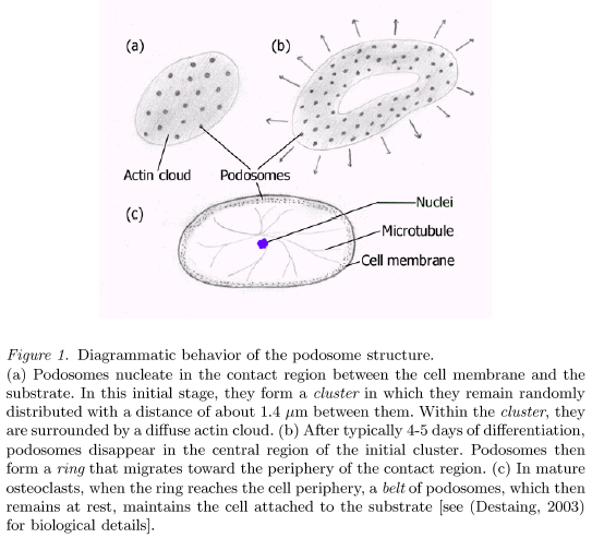
Podosomes appear like bright dots surrounded by a cloud in the microscope [fluorescence microscopy, the actin monomers (actin-GFP) are fluorescent]. They form three different collective structures depending on the considered stage of differentiation.
The model
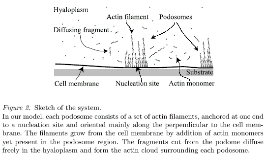
We consider podosomes as assemblies of actin filaments mainly oriented along the perpendicular to the cell membrane (Fig.2). The two basic ingredients of the model are a polymerization frequency v (a new actin monomer is attached to each filament with the typical frequency v) and a severing frequency b (each bond between two actin monomers within a filament is cut with the typical frequency b).
Results
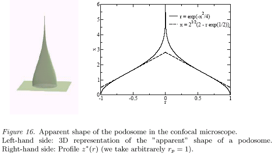
From our model, we recover the apparent shape of the podosomes in the confocal microscope (Fig.16). The height of the structure is predicted to scale like (v/b)1/2 whereas the characteristic time for the fluorescence recovery in the FRAP experiments is found to be about 1/(vb)1/2. The model also accounts for the characteristic size of the actin cloud that surrounds the podosome structure. Moreover, the model predicts that there exists a range of the overall monomeric-actin concentration in which the podosomes are expected to exhibit a steady shape for a finite life-span. This result is in agreement with the experimental observations.
Related publication
Podosomes
display actin turn-over and dynamic self-organization
in
osteoclasts expressing actin-GFP,
Destaing O. and Saltel F., Géminard J.-C., Jurdic P., and Bard
F.,
Mol. Biol.
Cell 14
(2003)
407-416.
Dynamics
of
bio-polymeric
brushes
growing
from a
cellular
membrane :
tentative
modelling
of the
actin
turnover
within an
adhesion
unit; the
podosome,
Biben T.,
Géminard
J.-C., and
Melo F., J. Biol. Phys., 31 (2005) 87-120.
Internal dynamics of actin structures involved in the cell
motility and adhesion: modeling of the podosomes at the molecular
level.
S. Hu, T. Biben, X. Wang, P. Jurdic and J.-C. Géminard,
Journal of Theoretical Biology 270, (2011) 25-30.